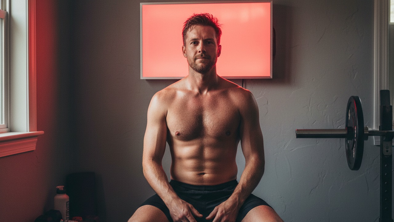
A dose of red light that helps a cell survive can, turned up just a little further, start killing it instead. That is the uncomfortable finding at the center of a new laboratory study on photobiomodulation, the clinical name for red and near-infrared light therapy, published in Photodiagnosis and Photodynamic Therapy in October 2026.
Testing light on the cells that build blood vessels
The researchers worked with human umbilical vein endothelial cells, known in lab shorthand as HUVECs. They are the standard stand-in scientists use to study the thin layer of cells that lines every blood vessel in the body.
That lining matters enormously for wound healing. Before skin can close over an injury, the body has to grow new capillaries into the wound bed to deliver oxygen and nutrients, a process called angiogenesis that depends entirely on endothelial cells like these.
The team exposed cultured HUVECs to varying parameters of red and near-infrared light, then measured two things: whether the cells survived and multiplied normally, and how quickly they closed a lab-made gap in a cell layer, a classic stand-in for how fast a wound bed migrates shut.
Low doses helped, higher doses did not
The pattern that emerged was not a straight line. Moderate light exposure improved both cell viability and the speed at which cells migrated across the gap, essentially accelerating the lab version of wound closure.
Push the exposure higher, though, and that advantage flattened out or reversed. This mirrors a long-standing observation in photobiomodulation research known as a biphasic dose response, where a therapeutic window exists and more energy past that window stops helping and can start to suppress cell function.
It is the same dose-response curve researchers have documented since early reviews of low-level laser therapy over a decade ago, and this study adds endothelial cells specifically to that evidence base.
Why light talks to mitochondria
The leading explanation for how red light affects a cell starts inside its mitochondria. Red and near-infrared wavelengths, generally in the range of 630 to 850 nanometers, are absorbed by an enzyme called cytochrome c oxidase, a key component of the mitochondrial chain that produces ATP, the cell’s energy currency.
Michael Hamblin, a photomedicine researcher who spent years at Massachusetts General Hospital and Harvard Medical School studying this pathway, has described cytochrome c oxidase as the likely photoacceptor that converts a dose of red light into a biological signal. Absorbing that light briefly displaces nitric oxide bound to the enzyme, which temporarily boosts ATP output and triggers a burst of reactive oxygen species.
In small amounts, that burst acts as a signal rather than damage. It switches on transcription factors tied to cell proliferation, migration, and the release of growth factors, which is plausibly why the cells in this study moved faster across the wound gap at the right dose.
Why endothelial cells specifically matter here
Praveen Arany, a researcher at the University at Buffalo who has spent years studying photobiomodulation’s effect on tissue repair, has focused much of his work on how light-triggered reactive oxygen species activate a dormant growth factor called TGF-beta1. That pathway helps coordinate the broader repair process, including the recruitment of new blood vessel growth into injured tissue.
Endothelial cells sit at the center of that recruitment. If photobiomodulation can nudge them to proliferate and migrate faster without pushing past the dose where benefits flip, it offers a plausible cellular explanation for why red light therapy has shown wound healing promise in some clinical and animal studies.
Studies in this area often pair a migration assay with a tube formation assay, where endothelial cells are placed on a gel-like matrix to see whether they organize into capillary-like networks, the functional signature of new blood vessel growth. Though this paper centers on viability and migration, those two measures are generally treated in the field as reasonable proxies for how well a given light dose supports the angiogenic side of wound repair.
That said, a petri dish is not a person. HUVEC experiments isolate one cell type under controlled lighting, stripped of the blood flow, immune activity, and tissue architecture present in a real wound.
From orbit to the living room
Photobiomodulation’s path into wellness panels did not start with influencers. NASA-funded research in the 1990s explored whether red and near-infrared LEDs could speed wound healing for astronauts, whose injuries tend to heal more slowly in microgravity.
That early work helped seed decades of clinical study into skin, muscle, and joint applications, and the same wavelength bands now show up in consumer panels sold for recovery and skincare.
The home device problem: more is not better
This is where the dose-response curve becomes a practical warning rather than just a lab curiosity. Red light panels marketed for skin and recovery encourage longer sessions and closer distances as a path to better results, but the biology described here argues against that instinct.
Devices like the one reviewed in Athletic Insight’s Hooga Pro panel test and the Joovv Solo 3.0 ship with manufacturer-specified distances and session lengths for a reason tied to this exact dose curve. Someone using a panel for a skin injury or for joint-focused protocols like those covered in Athletic Insight’s guide to red light therapy for knee pain is better served sticking to the tested distance and duration than maximizing exposure time in hopes of a bigger effect.
Underdosing simply wastes a session. Overdosing, based on this study and the broader dose-response literature it builds on, may actively work against the cells doing the repair.
What this does and does not prove
This is cell culture work, not a clinical trial, and HUVECs exposed to a lamp in a dish behave differently than skin exposed to a consumer panel at a distance through clothing or air. The specific energy levels that helped these cells will not map directly onto a home device’s settings without further research connecting the two.
Closing that gap will likely require in vivo wound models and eventually randomized trials on real skin injuries, where blood flow, immune cells, and skin pigmentation all influence how much light actually reaches the tissue underneath. Darker skin, for instance, absorbs and scatters more light before it reaches deeper structures, which complicates translating a lab dose into a universal device setting.
What it does reinforce is a theme that keeps surfacing across photobiomodulation research: the mechanism is real, measurable at the cellular and mitochondrial level, and bounded by a dose window rather than scaling endlessly with more light.
The appeal of a red light panel is that it feels like a dial you can simply turn up. This study is a reminder that the biology underneath it behaves more like a window you can walk past, not a volume knob you can max out.